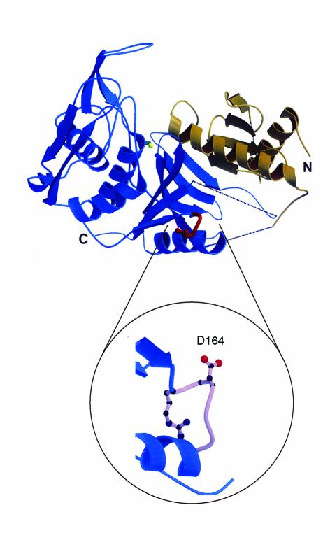
Figure 1.
Ribbon diagram of the three-dimensional structure of SpeB, an extracellular cysteine protease virulence factor. The protease domain is shown in blue, the prosegment in brown, the catalytic site cysteine in green, and the RGD loop embellishment in red. Dotted lines indicate flexible regions. The RGD loop is magnified to reveal the exposed Asp-164 residue required for binding of αvβ3 and αIIbβ3 human integrins. Serotype M1 strains that are abundant causes of human disease contain the RGD motif in the integrin-binding loop, whereas other strains possess an RSD sequence that does not bind integrins (8). N, amino-terminus; C, carboxy-terminus. Modified from ref. 23with permission.

