
Figure 1.
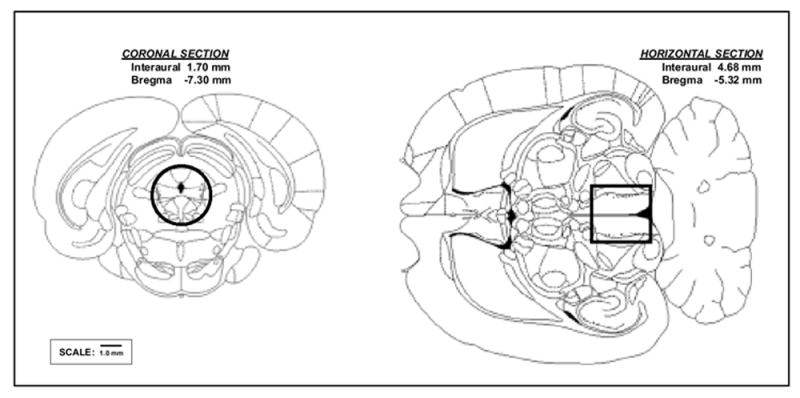
Reconstruction of the periaqueductal gray (PAG) tissue punch used in this study based on the atlas of Paxinos & Watson (1998). PAG was isolated by taking a circular punch (3 mm inner diameter) through the PAG from a transverse section of a coronal section of the brain cut from the anterior boundary of the superior colliculus to the posterior boundary of the junction between the inferior colliculus and the cerebellum.