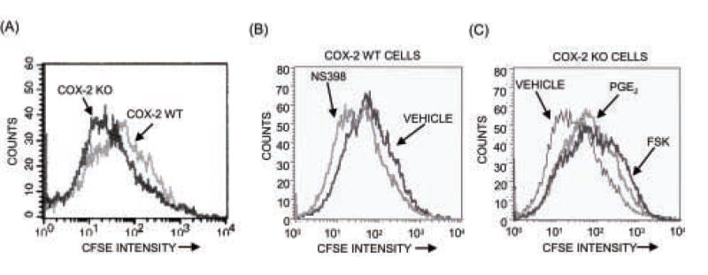
Figure 4. Assessment of cell division by CFSE staining.

Cells were stained with CFSE before plating, cultured for 7 days and analyzed by flow cytometry, as described in Materials and Methods. (A) Comparison of cell division in COX-2 WT and KO cells. (B) Effects of NS398 (0.1 μM) on cell division in COX-2 WT cells. (C) Effects of PGE2 (1 μM) or forskolin (10 μM) on cell division in COX-2 KO cultures.
