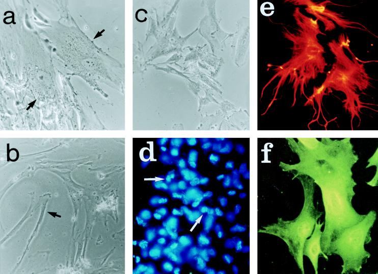
Figure 2.
Photomicrographs that demonstrate some of the morphological characteristics of MSCs and astrocytes in culture. a and b demonstrate two types of human MSCs, flat and elongated. c demonstrates the similar morphology of rat MSCs. d shows fluorescent labeling of nuclei of human MSCs immediately before implantation. e and f demonstrate indirect immunofluorescent staining of astrocytes with antibodies against vimentin and glial fibrillary acidic protein, respectively. (Magnification: a, b and c, ×20; d, ×10; e and f, ×40.)

