
Figure 5. Spontaneous Activity Confines GluR1 Movement Inside Synapses.
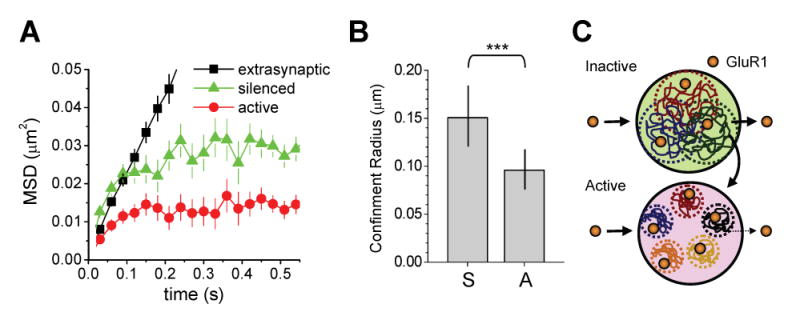
(A) Mean square displacement (MSD) versus time for GluR1-QDs in the indicated compartments. Extrasynaptic GluR1 undergoes free diffusion without confinement as indicated by the linear MSD curve. GluR1 receptors at synapses exhibit confined movement within a zone whose radius is defined by the maximum MSD value approached at the t = ∞ limit. Error bars indicate SD.
(B) GluR1 diffusion is more confined at active synapses. Data represent means ± SD of the confinement radius for GluR1 lateral movement in silenced (S) and active (A) synapses, as determined by the MSD curves in (A). Silenced, n = 125 trajectories reconstructed from 34 image fields on 13 coverslips. Active, n = 175 trajectories reconstructed from 26 image fields on 11 coverslips. ***p < 0.01, ANOVA.
(C) A schematic model for GluR1 lateral diffusion at active and inactive synapses viewed en face. Input-specific spontaneous synaptic activity reduces receptor mobility, limits exchange with the extrasynaptic membrane, and confines GluR1 within small subdomains of the postsynaptic membrane. This diffusional trap leads to GluR1 accumulation at active synapses. See text for details.