
Fig. 3. Fo-Fc difference map of (A) p- and (B) n-side binding site of TDS in the b6f complex.
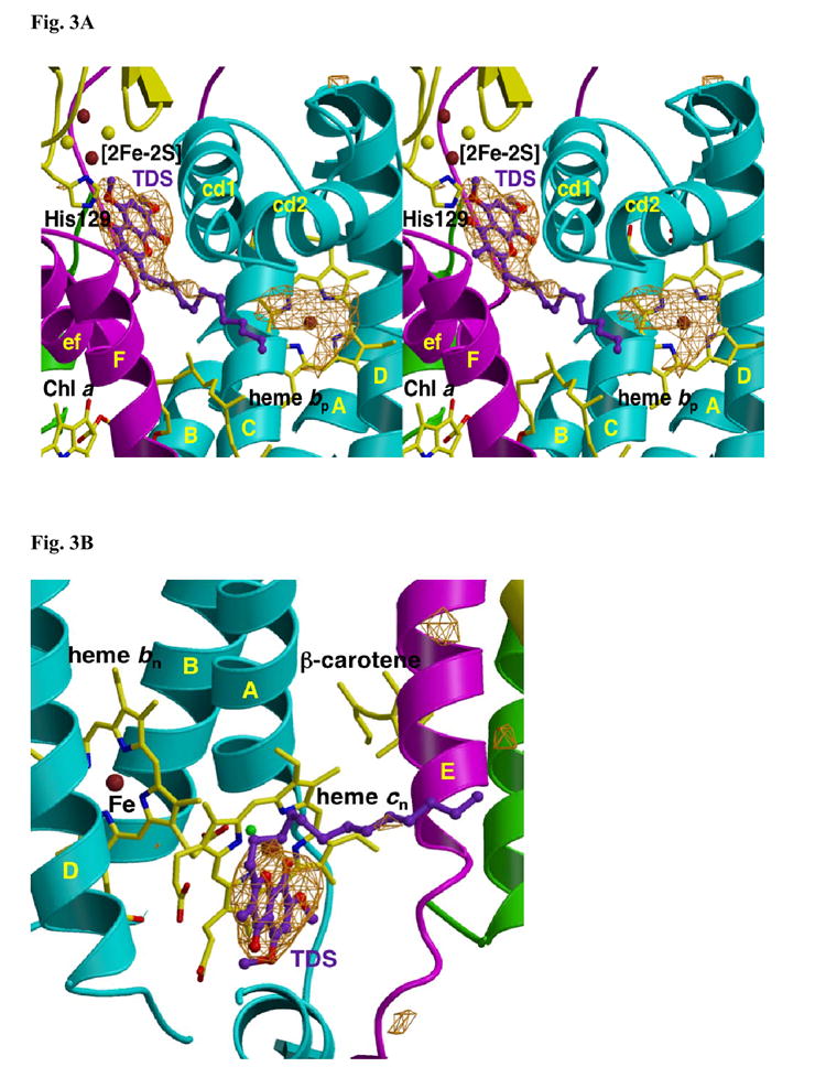
(A) p-side. The extra density within H-bond distance of the His129 ligand of the [2Fe-2S] cluster and between the ‘ef’ and ‘cd1’ loops is attributed to the chromone ring of TDS. As in Fig. 2A, the origin of the electron density under the cd2 loop, presumably lipid and/or detergent, is not known. (B) n-side. The position of TDS is shown relative to heme cn, which is exposed to the quinone-exchange cavity. TDS is on the side of heme cn distal to heme bn. TM helices and surface helices within loops labeled as in Fig. 2. Fo-Fc maps contoured at 4σ.