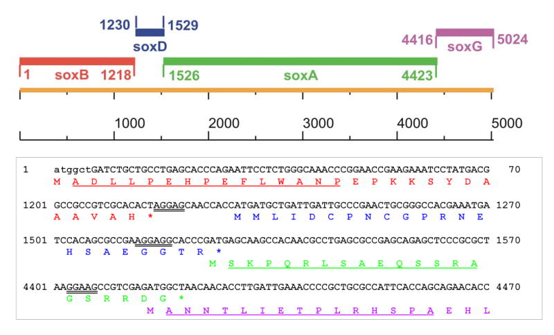
Figure 3.

The top panel shows the organization of the pTSOX genes. The bottom panel shows the nucleotide sequence at the 5′ end of each of the pTSOX genes and the intergenic regions along with the corresponding deduced amino acid sequence. The first 6 bp of soxB were missing in the original clone (pME4) and are shown in lower case. The deduced amino acid sequences of the β, δ, α and γ subunits are shown in colors (red, blue, green and magenta, respectively) that match those used for the corresponding genes in the top panel. Putative ribosomal-binding sites are indicated by double underlining. N-terminal peptide sequence data were obtained for all subunits except δ. The observed peptide sequences (indicated by single underlining) matched the deduced amino acid sequence.
