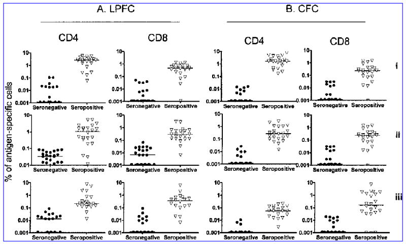
FIG. 2.

CD4+ and CD8+ T cell responses of CMV-seropositive and CMV-seronegative donors to stimulation with CMV lysate (i), pp65 peptide pool (ii), and IE peptide pool (iii). CD4+ and CD8+ T cell proliferation (A) was significantly higher in the CMV-seropositive donors, compared with the CMV-seronegatives for all three antigens (p < 0.0001 for all comparisons except CD8+ IE response, p = 0.0005). Likewise CD4+ and CD8+ T cell IFNγ expression, measured in the CFC assay (B), was significantly higher in the CMV-seropositives (p < 0.0001). IFNγ expression and proliferation were quantified as a percentage of the total CD4+ (gated on CD3+CD4+) or CD8+ (gated on CD3+CD4−) T cell population.
