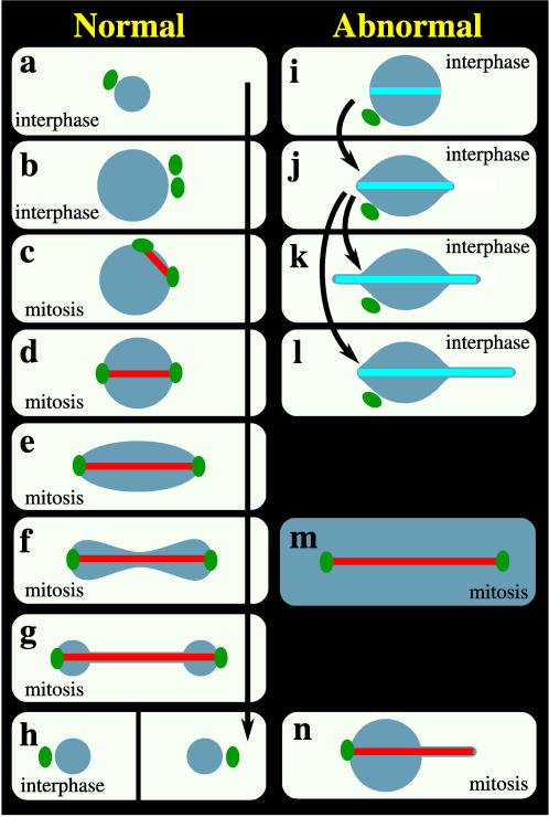
Figure 1. Normal nuclear geometric transformations in the life cycle of fission yeast and a catalogue of fission yeast nuclear abnormalities.
In interphase (a to b), which lasts for 3–4 hr, the volume of the nucleus increases with time to twice the initial volume of ∼4π/3×1.13 µm3. When mitosis begins, the duplicated SPBs (b) are embedded in the NE (c) and as they assume positions on opposite sides of the nucleus (c, d) nucleate the assembly of MTs that form the mitotic spindle. As the spindle elongates (d–f) to a final length of 12–15 µm [15], the initially spherical nucleus (d), measuring ∼1.1×21/3 µm in radius, is deformed consecutively into an oval (e), peanut (f), and dumbbell (g) shape before resolving into two spherical daughter nuclei (h) ∼1.1 µm in radius. Cytokinesis (h) then physically separates the nuclei into two individual cells (a) that initiate another round of cell division. (i to l) Formation and elongation of a n-MTB in interphase cells, leading to formation of one or two tethers, upon ned1 overexpression [6]. (m) NE fragmentation during mitosis, in strains in which the Ran GTPase system is perturbed [reviewed in 35]. (n) During abnormal mitosis in the cut11-2 temperature sensitive mutant [11], the msd1 null mutant [12] or upon mia1 overexpression [13] or laser microsurgery [14], one end of the mitotic spindle has no SPB or is not properly anchored to the SPB and, upon spindle elongation, this end induces a single tether whereas the NE appears undeformed at the other end. Dark blue: nucleus; green: SPB; red: spindle in mitotic cells; turquoise: n-MTB in interphase cells.

