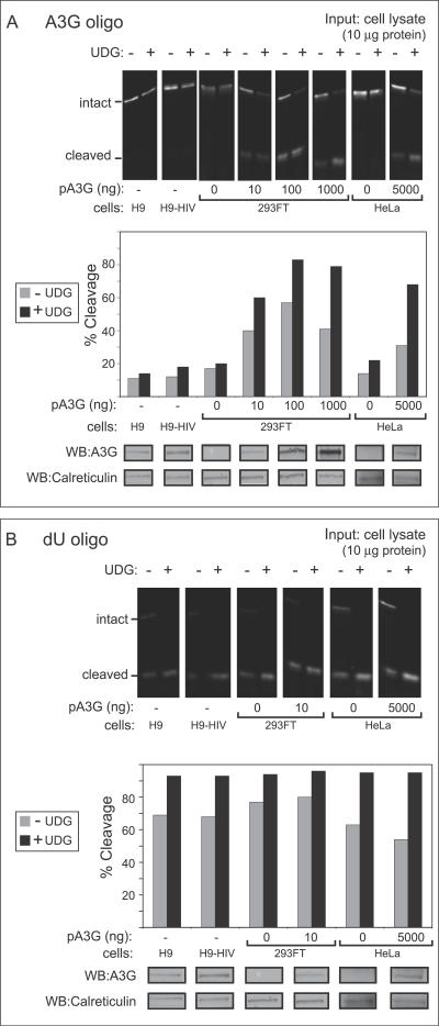
Figure 1. A Gel-Based Assay Reveals That Endogenous A3G in T Cell Lines Exhibits Unexpectedly Low Deaminase Activity Compared to Exogenous A3G in Transfected Epithelial-Derived Cell Lines.
(A) Deaminase activity was measured using an infrared 700 (IR700)–labeled oligo containing the A3G recognition site (CCC) either with or without exogenous recombinant uracil DNA glycosylase (+/- UDG). Oligos were incubated with crude cell lysates containing 10 μg of total cellular protein obtained from H9 cells, H9 cells expressing the HIV genome containing a deletion in Vif (H9-HIV), or from HeLa or 293FT cells transfected with the indicated amounts of A3G plasmid DNA (pA3G). Extent of oligo cleavage (indicating extent of deamination) was determined by gel electrophoresis followed by detection on a LI-COR scanner (top panel), and the percentage of probe cleaved was graphed (second panel). Below, equivalent amounts of cell lysate were analyzed in parallel by western blot (WB) to show A3G protein content. Western blot of calreticulin is shown as a loading control.
(B) UDG activity was measured in select lysates from (A) using an IR700-labeled dU-containing oligo in the presence or absence of exogenous UDG (+/- UDG). Results are displayed as in (A) and show that unlike A3G activity shown in (A), UDG activity is similar in all cell lysates analyzed.
All assays were performed on RNAse A–treated samples.

