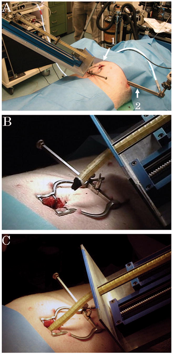
FIGURE 2.

A, The robot is mounted on the table. A small incision is made at the subxiphoid (arrow 1). Visualization is achieved by a thoracoscope inserted from the left thoracic wall (arrow 2). B and C, The robot advances forward into the intrapericardial space through the subxiphoid incision.
