
Figure 7.
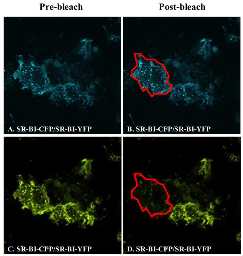
Photobleaching of YFP fluorescence in COS-7 cells expressing SR-BI-CFP/SR-BI-YFP constructs. COS-7 cells were transiently co-transfected with SR-BI-CFP and SR-BI-YFP proteins. Panels A, B show CFP fluorescence following excitation at 458 nm. Panels C, D show YFP fluorescence following excitation at 514 nm. Panels A, C: images taken prior to acceptor photobleaching; panels B, D: images taken after acceptor photobleaching. Images are representative of approximately 10–18 images taken for this particular experimental condition, which was performed at least 4 times. The red outline indicates the region of interest chosen for FRET measurements.