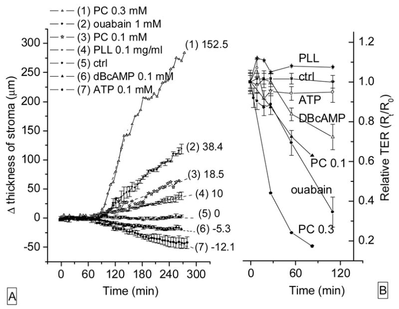
Figure 11.

Effects of agents on stromal thickness and relative electrical resistance. (A): rabbit corneal endothelial preparations. Numbers to the right identify the curves and represent rates (μm/h) of swelling of corneal stroma for 60 minutes after drug application. (B): TER across CRCEC layers on permeable supports challenged with the same compounds used in the left (A) panel. Data represent average ± S.E.
