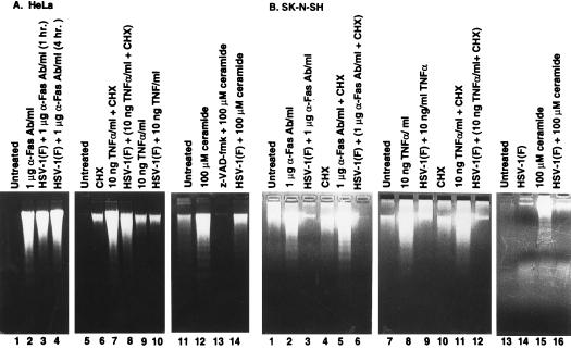
Figure 3.
Photograph of agarose gels containing electrophoretically separated low molecular weight DNA fractions from mock-infected or infected cells and stained with ethidium bromide. Subconfluent HeLa (A) or SK-N-SH cells (B) were mock-infected or infected with HSV-1(F) and treated by incubation in 1 μg/ml of anti-Fas IgM or 10 ng/ml TNFα both in the presence or the absence of 1 μg/ml CHX. Anti-Fas IgM or TNFα-treated HeLa cells were collected at 8 and 6 hr after treatment, respectively. The SK-N-SH cell samples were collected at 12 and 16 hr after treatment, respectively. Mock-infected and infected HeLa and SK-N-SH cells were treated with 100 μM C2-ceramide and collected at 6 hr after treatment.

