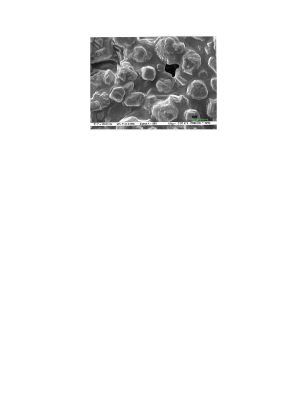
Figure 3.
Scanning electron microscopy (SEM) image of CLEA: SEM was carried out on Zeiss EVO50 scanning electron microscope, UK. Sample was dried by rinsing with anhydrous acetone, placed on a sample holder, coated with silver before being scanned under vacuum. The particle size was determined from the micrograph with the scale of 20 μm unit.

