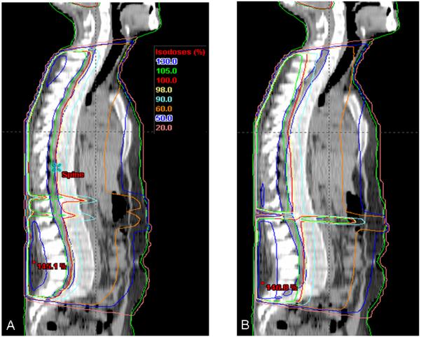
Figure 5. Comparison of mid-sagittal isodose curves for plans requiring matched PA spinal fields.
Mid-sagittal isodose curves are pictured from the same patient planned with two IMRT spine fields (A) and two conventional spine fields (B). Note the improved coverage of the cervical and lumbosacral spine with the IMRT plan compared to the conventional plan.

