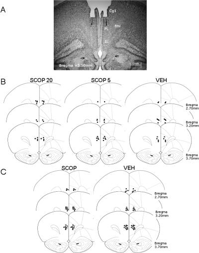
Figure 1.
(A) Photomicrograph of Cresyl violet staining at the level of the PL area (AP, 3.50 mm anterior to bregma) showing the cannula track and the microinjector tip of a representative subject. (B,C) Microinjector tip placements for different groups throughout the rostral-caudal extent of the PL (from 3.70 to 2.70 mm anterior to bregma) in experiments 1 (B) and 2 (C). Reprinted with permission from Elsevier © 1997, Paxinos and Watson (1997). (●) n = 1, (*) n = 2, (+) n = 3. (Cg1) Cingulated cortex area 1; (fmi) forceps minor of the corpus callosum; (IL) infralimbic cortex; (PL) prelimbic cortex.

