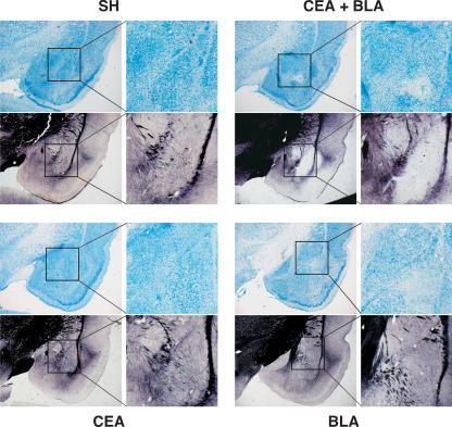
Figure 4.
Representative slices stained with thionin and AuCl. Slices shown from rats that received lesions of the CEA + BLA (upper right), CEA (lower left), or BLA (lower right); SH rats (upper left). Both the thionin- and the AuCl-stained slices for each group are taken from the same representative animal at approximately the same anterior–posterior level with a magnification of the amygdala shown to the immediate right.

