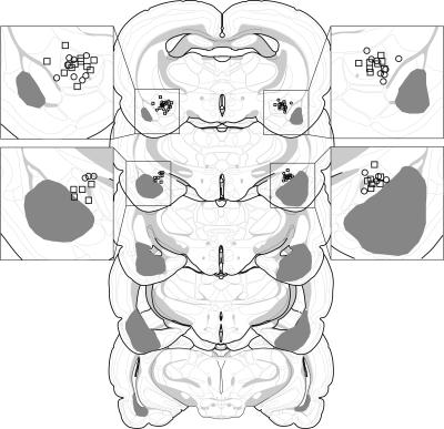
Figure 9.
Schematic representation of the extent of pretraining NMDA lesions (median lesion) of the BLA (dark gray) and the locations of included cannula placements for the infusion of muscimol (circles) or 0.9% sterile saline (squares) in the CEA (Experiment 4). A magnification of the amygdala is shown adjacent to the coronal brain sections. Coronal brain section images adapted from Swanson (1992).

