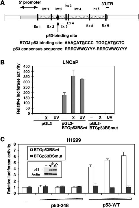
Figure 2.
DNA damage-induced expression of BTG3 is mediated through a putative p53-binding site located in intron 2. (A) Schematic diagram of BTG3 including the 5′ promoter region, exons (Ex) and the neighboring introns (Int). The p53 consensus binding site is marked and indicated by an arrow. Also shown is the comparison of the BTG3 p53-binding site with the consensus sequence known for p53 binding. There is no spacing between the two half sites in the BTG3 p53 binding sequence. (B) The putative p53 binding sequence in intron 2 can mediate the DNA damage response independent of other intron sequences. Luciferase assays were performed with a reporter driven by the p53 binding sequence isolated from intron 2 (pGL3-BTGp53BS). A reporter with mutated sequence (pGL3-BTGp53BSmut) was also examined. (C) The WT but not the mutated p53 binding sequence is activated by exogenously expressed WT p53 but not by the 248 mutant. All luciferase activities were presented after normalization to the cotransfected β-galactosidase control. Error bars represent standard deviations of at least three independent experiments.

