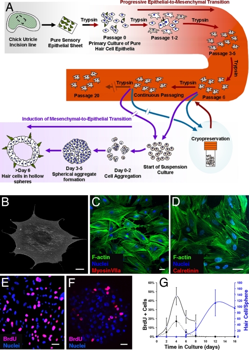
Fig. 1.
Advanced-passage cultures from pure hair cell epithelia. (A) A schematic diagram showing how inner-ear hair cells were produced in vitro. (B) Cells cultured from embryonic day 14 chicken embryo hair cell epithelia changed from columnar to flattened polygonal shapes as illustrated in this scanning electron micrograph of a cell from passage 3. (C and D) By passage 3, preexisting hair cells and the hair cell markers myosin VIIa and calretinin were no longer detectable in the 2D cultures, and phalloidin-labeled F-actin became organized in stress fibers in the cultured cells. (E–G) Cells in 2D cultures proliferated and continued to incorporate BrdU during 24-h labeling periods that preceded fixations at different stages (E and solid dark line in G), but the incidence of proliferation decreased after cells were suspended and formed spheres (F and dashed dark line in G, mean ± standard deviation). Hair cells differentiated as early as day 6 after suspension and increased in number for at least 6 more days (blue line in G). (Scale bars: 20 μm.)

