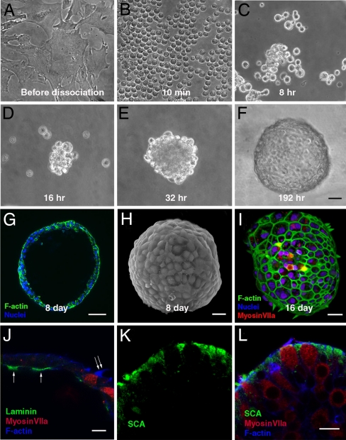
Fig. 3.
Aggregation of late-passage cells led to formation of hollow smooth-surfaced spheres with apical structures pointing outward. (A) Advanced passage cells growing in 2D culture. (B) A fully dissociated sample of cells 10 min after it was trypsinized from a 2D culture such as that in A and placed in suspension. (C–E) Cells adhered to each other and progressively formed larger aggregates at 8 (C), 16 (D), and 32 (E) h after dissociation from the 2D cultures and suspension in the serum-free medium. (F) The aggregates formed smooth spheres by day 8 after suspension. (G and H) An optical section showing the hollow lumen of such a sphere (G) and a scanning electron micrograph showing the characteristic smooth surface of a sphere 8 days after suspension of the cells from an advanced-passage 2D culture (H). (I) Phalloidin-labeled F-actin in hair bundles projecting outward from cells that are also labeled by anti-myosin VIIa. F-actin juxtaposed to epithelial junctions is also visible in this 3D reconstruction from confocal microscopy. (J) Laminin (arrows) is expressed in patches along the inner basal epithelial surface of the sphere shown and was found in similar patches in four other spheres. Note the hair bundle extending from the sphere's outer surface (double arrow). Other cells in the epithelia of the spheres express the supporting cell marker, SCA (in K and the merged image in L). (Scale bars: 20 μm in A–F; 50 μm in G; 30 μm in H and I; and 10 μm in J–L.)

