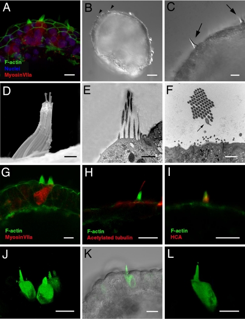
Fig. 4.
Bona fide hair cells form sensory hair bundles that project outward from the spheres. (A) Hair bundles on the apical surfaces of myosin VIIa-positive hair cells surrounded by epithelial junctions. (B and C) Hair bundles (arrowheads and arrows) at the surface of a sphere visualized by differential interference contrast microscopy. (D) Another hair bundle visualized by scanning EM. The spheres in B–D were produced from cells that had been cultured through 19 passages. (E and F) Longitudinal (E) and transverse (F) sections through hair bundles at the surfaces of spheres viewed by transmission EM. Note the characteristic eccentrically positioned kinocilium (arrow in F, which was observed in a fortuitous transverse section through one of the 18 hair bundles examined in the five spheres that were processed for transmission EM). (G) Phalloidin-labeled stereocilia on the surface of a myosin VIIa-positive hair cell and its neighbor, and F-actin outlined the surrounding columnar cells of the sphere's wall. (H) Anti-acetylated tubulin labeled a single kinocilium extending above the phalloidin-labeled hair bundles on at least 8 of the 21 myosin VIIa-positive hair cells observed in limited examinations of the five spheres immunostained with that antibody. (I) Hair bundle double-labeled with phalloidin and antibody to the 275-kDa hair cell antigen (HCA). (J–L) In optical assays for open mechanotransduction channels, FM1-43 permeated hair cells after 10-sec exposures to the dye that were followed by thorough rinsing, but no other cells in the spheres were labeled. The spheres and hair cells in this figure were all derived from cultures that had been frozen at P5, and then thawed and continuously cultured until P19, when they were dissociated and aggregated in suspension. (Scale bars: 10 μm in A, C, and G–L; 50 μm in B; and 1 μm in D–F).

