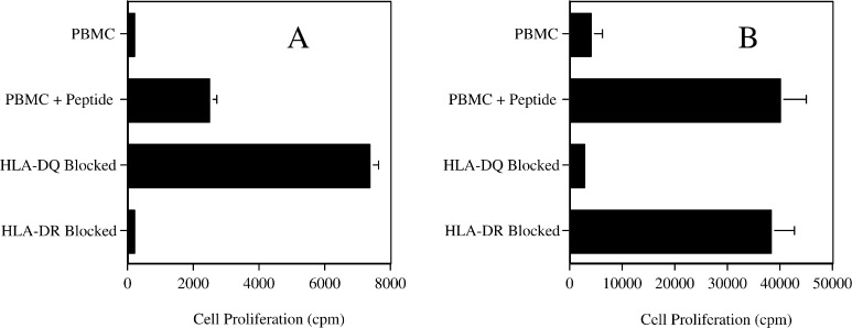
Fig. 2.
MHC restriction analysis of two peptide PAX3/FKHR-381 specific HTL lines (clone 5B in frame a and clone 9E in frame b) from different donors. Monoclonal antibodies specific for HLA-DR or HLA-DQ at 10 μg/ml were cocultured with peptide-pulsed (5 μg/ml) irradiated autologous PBMC and HTL. HTL proliferation was determined using the same culture conditions as described in Fig. 1. Proliferation was observed in response to peptide (PBMC + peptide) but not in the absence of peptide (PBMC). Values represent mean; bars SD.

