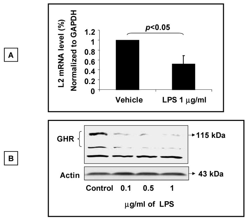
Fig.3. LPS inhibits expression of endogenous GHR.

(A) 3T3-F442A preadipocytes were exposed to LPS (1 μg/ml) for 5-6 h and cells harvested for extraction of total RNA. The steady state abundance of the L2 mRNA transcript of the GHR was measured by Real Time RT-PCR analysis. Expression of the housekeeping gene GAPDH was used as internal control to normalize the results. Results are expressed as mean ± SEM; n=3. p < 0.05 (Mann-Whitney U test) compared to absence of LPS (vehicle). (B) Analysis of GHR protein expression. 3T3-F442A preadipocyte cells were stimulated with LPS (1 μg/ml) for 5-6 h and whole cell lysates prepared. Aliquots of equal amounts of protein were size-fractionated by electrophoresis, transferred on to nitrocellulose membrane by Western blotting, and the membrane probed with antibody specific for the GHR (AL-47). Following detection of the GHR using the chemiluminescence system as described under “Experimental Procedures, the blot was stripped and reprobed for actin. The positions of the molecular weight markers, the GHR, non-specific band (NS), and actin are indicated. Results depicted are representative of two such experiments.
