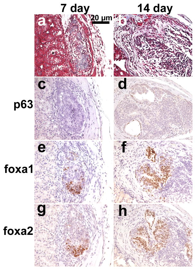
Figure 4.

Xenografts of 1500 embryonic stem cells + 4 embryonic bladder mesenchymal shells/graft harvested at 7 (a,c,e,g) and 14 (b,d,f,h) days. a&b. Gomori’s trichrome, cellular organization occurring in cells that look destined to become urothelium (blue). c&d. Immunohistochemical detection of p63 (brown) illustrating lack of expression within 7 day tissues and early expression observed in 14 day tissues. e&f. Immunohistochemical detection of Foxa1 (brown) denoting endodermal cells. g&h. Immunohistochemical detection of Foxa2 (brown) denoting endodermal cells.
