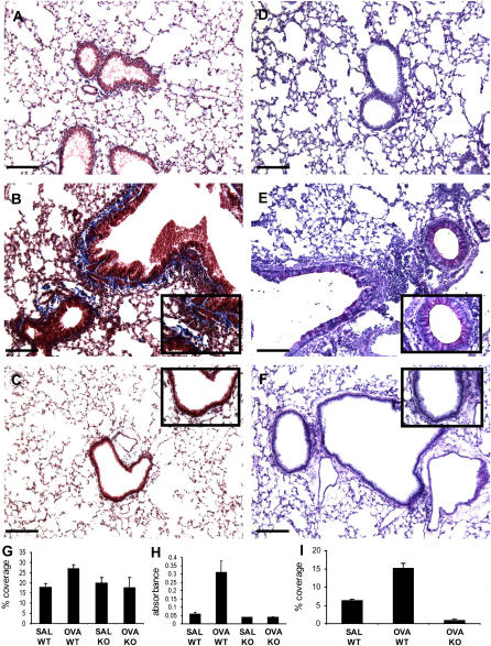
Figure 4.
Effects of phosphate-buffered saline (PBS) and ovalbumin (OVA) 3 hours after challenge on Day 21 in the lungs of Plg+/+ and Plg−/− mice. Representative photomicrographs of Masson trichrome (A–C)– and periodic acid-Schiff (PAS) (D–F)–stained lung sections are shown; n = 3–9 mice/group. PBS treatment had no effect on the lungs of Plg+/+ mice (A and D). OVA treatment resulted in increased deposition of collagen (blue) in large portions of the peribronchial area (B) as well as increased goblet cells and mucus production (magenta) in the bronchial epithelium (E) of Plg+/+ mice. These changes were not observed in the lungs of OVA-treated Plg−/− mice (C and F, respectively). Original magnifications for A–F are ×100. Higher magnification (×400) insets in B versus C and E versus F highlight the difference in collagen deposition and goblet cells and mucus production, respectively, between the genotypes. Bars = 137 μm. (G) Quantification of Masson trichrome–stained lung sections revealed a statistically significant increase (p = 0.009) in collagen deposition in OVA-treated Plg+/+ mice (n = 20) over their PBS counterparts (n = 11) and the PBS- (n = 9, p = 0.01) or OVA-treated Plg−/− mice (n = 10, p = 0.05). (H) Sircol assay using lung homogenates also showed a significant increase of new collagen in OVA-treated Plg+/+ mice (n = 4) over their PBS counterparts (n = 3, p = 0.05) and the PBS- (n = 3, p = 0.04) or OVA-treated Plg−/− mice (n = 3, p = 0.01). (I) Quantification of PAS-stained lungs sections revealed a significant increase of mucus production in OVA-treated Plg+/+ mice (n = 29) over their PBS counterparts (n = 16, p = 0.0001) and OVA-treated Plg−/− mice (n = 16, p = 0.001). SAL = PBS-treated; WT = wild-type.

