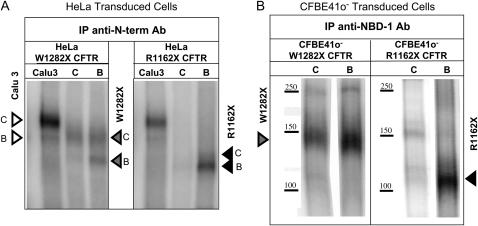
Figure 5.
Detection of truncated R1162X and W1282X CFTR in HeLa (A) and CFBE41o− (B) cells. (A) HeLa Cells were selectively probed for truncated CFTR using an antibody directed against NBD-1 in W1282X and R1162X CFTR–transduced cells. Truncated protein expression (core and fully glycosylated CFTR represented by open arrowheads) was increased after 24 h exposure to NaBu (lane B, 500 μM) compared with control conditions (lane C). Calu-3 cells are also shown as a control for wt protein expression. (B) Similar analysis in CFBE41o− cells. NaBu treatment significantly enhanced W1282X (∼ 140 kD, left panel) or R1162X (∼ 110 kD, right panel) CFTR protein expression. The absence of identifiable band C likely reflect more stringent control of glycosylation in the polarizing cell type (32).

