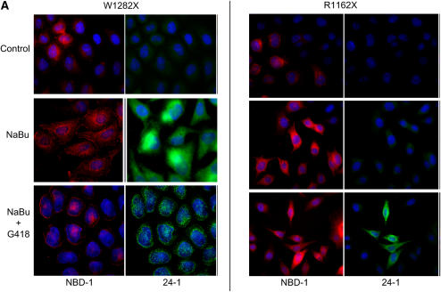
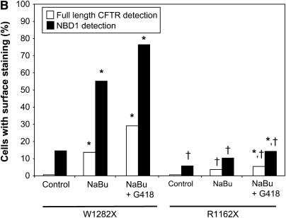
Figure 7.
NaBu increases surface-localized truncated CFTR expression in W1282X CFTR–expressing cells as detected by immunofluorescence. (A) Immunofluorescence staining of W1282X (left) and R1162X CFTR (right)–expressing HeLa cells after 24 h exposure to NaBu (2.5mM), NaBu + G418 (500 μg/ml), or untreated (vehicle control) conditions. Staining with anti–NBD-1 antibody for truncated CFTR is noted in red, while selective full-length CFTR staining is noted in green (anti–C-terminus antibody, 24–1). W1282X CFTR–transduced cells demonstrate significantly greater number of cells with surface localized truncated CFTR after 24 h treatment with NaBu compared with R1162X cells (55.2% versus 10.4% of cells with surface staining, respectively; P < 0.001). A significant number of cells with surface localized full-length CFTR is detected in both cell lines after the addition of G418, although a greater percentage was seen in W1282X-transduced cells (29.1% of W1282X cells exhibit surface staining after G418 + NaBu versus 0.5% under control conditions, P < 0.001; 5.4% and 0.06% surface staining was seen in R1162X cells, respectively, P < 0.01). (B) Summary figure representing percentage of cells with surface staining evident. Open bars represent full-length CFTR detection, and solid bars represent detection of CFTR with anti–NBD-1 antibody (truncated protein). Between 160 and 190 cells were scored for CFTR surface staining per treatment condition, and totaled over three separate experiments. *P < 0.05 versus control condition for each cell type; †P < 0.001 comparing W1282X versus R1162X cells at exact same treatment condition.


