
Fig. 6.
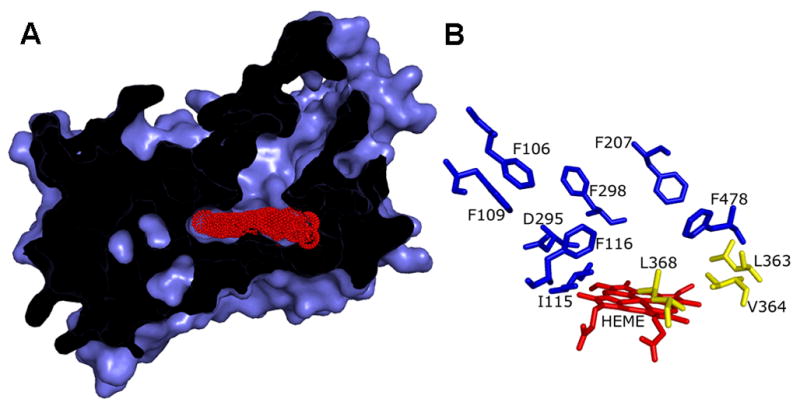
CYP2E1 homology model. (A) View shows the narrow channel leading from the solvent to the small active site. Access to the heme (red) by the substrate is partially occluded by the structure of the protein. (B) Diagram indicates CYP2E1 residues, which may interact with AzMC based on the alignment with CYP2A6 amino acids binding coumarin (Table 3). Residues lining the active site proximal to the heme and were potential targets for nMC incorporation are colored yellow. Molecular graphics were generated by PYMOL (http://pymol.sourceforge.net).