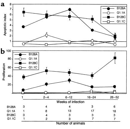
Figure 2.

H. pylori strains differentially alter gastric epithelial cell apoptosis (a) and proliferation (b) in Mongolian gerbil gastric mucosa. Apoptosis and proliferation scores at each time point are expressed as mean number of positive cells per gland in infected gerbils divided by mean scores in uninfected gerbils. Therefore, a value of 1 represents baseline. (a) Apoptosis scores were significantly higher in animals infected with GU strain B128 in the antrum (B128A; filled circles) and corpus (B128C; filled squares) 1–24 weeks after challenge compared with DU strain G1.1 (open symbols) or controls (P = 0.01 and 0.009, respectively); scores subsequently decreased, and by 26–52 weeks, they were not different from controls. Compared with uninfected animals, apoptosis was also significantly increased in gerbils infected with DU strain G1.1 but only in the antrum (G1.1A, open circles) and only at 2–4 weeks after inoculation (P = 0.01). (b) Proliferation scores increased shortly following challenge with GU strain B128 (filled symbols) and were significantly higher at all time points than either G1.1-infected (open symbols) or control animals (P < 0.001 for each).
