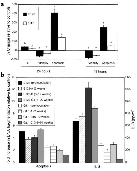
Figure 3.

Quantitation of IL-8 and analysis of apoptosis among preinoculation (parental) strains B128 and G1.1 (a) and their derivative progeny after gerbil passage (b). (a) AGS cells were grown alone or in the presence of H. pylori GU strain B128 (filled columns) or DU strain G1.1 (open columns). IL-8 concentrations were determined 24 hours after inoculation by ELISA. No 48-hour samples were analyzed due to the significant loss of AGS cell viability that had occurred by this time point. Cell viability and apoptosis were assessed by trypan blue exclusion and DNA fragmentation ELISA, respectively, 24 and 48 hours following inoculation. Results are expressed as the percent change in IL-8, viability, or apoptosis relative to controls. Data represent mean ± SD of three independent experiments. AP ≤ 0.05 compared with AGS cells alone. BP ≤ 0.05 compared with controls or with DU strain G1.1. (b) Six gerbil-passaged B128 and G1.1 isolates were selected (B128-A and G1.1-A, 2 weeks; B128-B and G1.1-B, 6–12 weeks; B128-C and G1.1-C, 16–20 weeks after challenge, respectively). Apoptosis results are expressed as levels of nucleosomal release relative to controls. IL-8 concentrations are expressed as pg/ml. Bars, SD.
