
Figure 2.
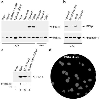
Expression of IRE1β is restricted to the epithelium of the gastrointestinal tract. (a) Immunoprecipitation (IP) followed by immunoblot detection of IRE1β and IRE1α in tissue extracts from wild-type and IRE1β–/– mice. (b) Detection by immunoblotting of IRE1β in microsomes prepared from all three segments of the gastrointestinal mucosa and pancreas. The ER resident protein ribophorin I, detected by immunoblot, serves as a recovery marker. (c) Detection of IRE1β by immunoprecipitation and immunoblotting in lysates prepared from epithelial cells eluted by EDTA treatment from the colon of a wild-type mouse. Lanes 1 and 2 are from lysates prepared from the intact tissue. Lane 3 is from the cells eluted from the tissue fragment in the calcium-free EDTA-containing buffer, and lane 4 is from a lysate prepared from cells that remain associated with the tissue fragment after (partial) depletion of epithelial cells. Lane 1 was immunoprecipitated with preimmune serum (PI), whereas lanes 2–4 were immunoprecipitated with anti-IRE1β immune serum. (d) Immunostaining of cells in an aliquot of the fraction used in lane 3 of c, with mAb’s to keratins. A counterstain with the karyophilic dye H33258 reveals the nuclei of all the cells in the field. Note that almost all the cells in the field stained positive with the keratin antibodies, attesting to their epithelial identity.