
Figure 4.
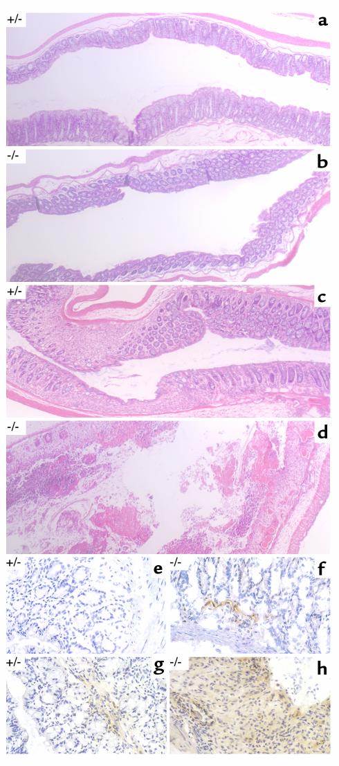
Histopathology of DSS-treated mice. Representative photomicrographs (×50) of paraffin-embedded, hematoxylin and eosin–stained longitudinal sections of the distal colon from untreated mice of the indicated genotypes (a and b) or mice that had been exposed continuously to 2% DSS for 12 days (c and d). Photomicrographs (×400) of longitudinal frozen sections of colon stained with an mAb to murine ICAM-1 revealed by diaminobenzidine (brown precipitate) and lightly counterstained with hematoxylin for orientation. Mice of the indicated genotypes were exposed to 2% DSS for 6 days (e and f) or 10 days (g and h).