
Figure 2.
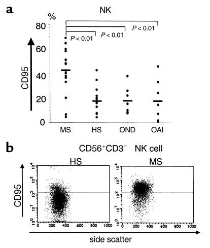
Surface expression of CD95 on NK cells in MS-rem and controls. (a) Frequency of CD95+ NK cells. Freshly isolated PBMCs were stained with anti–CD3-FITC, anti–CD56-PE, and anti–CD95-biotin/Streptavidin-Cychrome and were analyzed by flow fluorocytometry within 2 hours after drawing blood. The data are expressed as percentages of CD95+ cells among CD56+CD3–-gated NK cells. Controls for MS-rem include HS, patients with other neurological diseases (OND) (Parkinson disease [n = 4], spinocerebellar degeneration [n = 2], amyotrophic lateral sclerosis [n = 1]) and those with other autoimmune diseases (OAI) (thyroiditis [n = 5], myasthenia gravis [n = 1], inflammatory demyelinating polyneuropathy [n = 1]). Kruskal-Wallis test with Scheffe’s F test was used for statistical analysis. Horizontal bars indicate mean values. (b) CD95 expression on NK cells from MS-rem versus HS. PBMCs were stained with anti–CD3-FITC, anti–CD56-PE, and anti–CD95-biotin/Streptavidin-Cychrome. CD56+CD3– lymphocytes were gated and analyzed for CD95 expression versus side scatter. The horizontal bars indicate the fluorescence intensity distinguishing CD95+ and CD95– cells. Shown are representative data out of 20 samples (MS [n = 10], HS [n = 10]) with similar results.