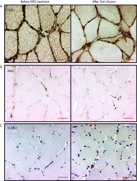
Figure 2 (A) Major histocompatibility complex class I antigen expression on the muscle fibre membrane and sarcoplasma of the muscle fibres (brown staining) before intravenous immunoglobulin (IVIG) treatment remained unchanged in the repeat biopsies (patient 12, inclusion body myositis). (B) Membranolytic attack complex (MAC) deposits in the capillaries (arrows, brown staining) before IVIG treatment remained unchanged in the repeat biopsies (patient 6, polymyositis). (C) Intercellular adhesion molecule‐1 (ICAM‐1) expression in capillaries (arrow, red staining) and inflammatory cells remained unchanged in the repeat biopsies (patient 9, dermatomyositis). Scale bar = 50 μm (original magnification ×250).

An official website of the United States government
Here's how you know
Official websites use .gov
A
.gov website belongs to an official
government organization in the United States.
Secure .gov websites use HTTPS
A lock (
) or https:// means you've safely
connected to the .gov website. Share sensitive
information only on official, secure websites.
