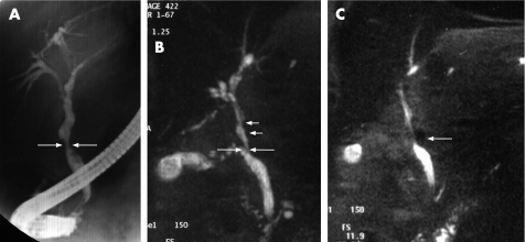
Figure 3 (A) Endoscopic retrograde cholangiogram in a patient with extrahepatic portal venous obstruction showing irregular common hepatic and intrahepatic bile ducts with smooth narrowing of middle common bile duct (CBD; arrows). (B) Magnetic resonance (MR) cholangiography of the same patient showing narrowing of middle CBD (long arrows) with indentations above it (small arrows). (C) MR cholangiography also shows the close association between narrowing of CBD and a collateral vein (arrow) with dilatation of intrahepatic ducts.

An official website of the United States government
Here's how you know
Official websites use .gov
A
.gov website belongs to an official
government organization in the United States.
Secure .gov websites use HTTPS
A lock (
) or https:// means you've safely
connected to the .gov website. Share sensitive
information only on official, secure websites.
