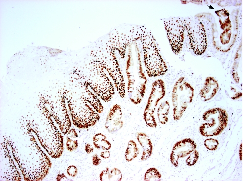
Figure 12 Native Barrett's glandular tissues stained with the proliferation protein Minichromosome maintenance protein 2 and counterstained with haematoxylin, demonstrating oesophageal glands fusing with the squamous epithelial surface. A segment of columnar epithelium on the far right contains glandular debris, which could result from the fusion of glands with the new epithelial surface (arrow) (×200).

An official website of the United States government
Here's how you know
Official websites use .gov
A
.gov website belongs to an official
government organization in the United States.
Secure .gov websites use HTTPS
A lock (
) or https:// means you've safely
connected to the .gov website. Share sensitive
information only on official, secure websites.
