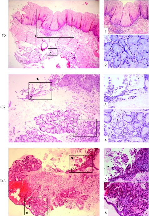
Figure 2 Squamous oesophageal endoscopic biopsy specimens were cultured with 100 μM all‐trans retinoic acid ATRA ex vivo over a 48 h time period (T). These images are representative sections, always orientated in relation to the biopsy surface (top) and stained with H&E. The images in the left‐hand side panel are at ×40 (T0, T48) and ×100 (T32) magnification and the images on the right corresponding to the numbered boxes are at ×100 (1), ×200 (5, 6) and ×400 (2–4) magnification. (T0, prior to culture): native squamous oesophageal mucosa with stratified squamous epithelium, lamina propria containing connective tissue or stroma (box 1), the muscularis mucosa and the submucosal glands (box 2) prior to culture. T32, (32 h of culture): the squamous mucosa is sloughing off from the surface (box 3, arrowhead) and the submucosal glands are of varying degrees of differentiation with some enlargement and rounding‐up of nuclei (box 4). T48, (48 h of culture): by 32 and 48 h of culture, the squamous mucosa has mainly sloughed off (arrowhead), leaving the basal layer (box 5) and glands seen cupping and fusing with the surface, which, in places, has a columnar‐lined appearance with a single layer of epithelial cells (arrow, box 6).

An official website of the United States government
Here's how you know
Official websites use .gov
A
.gov website belongs to an official
government organization in the United States.
Secure .gov websites use HTTPS
A lock (
) or https:// means you've safely
connected to the .gov website. Share sensitive
information only on official, secure websites.
