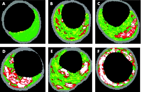
Figure 3 Plaque classification by intravascular ultrasound–virtual histology distinguishes between intimal thickening (A, B), and more vulnerable lesions, such as fibroatheroma (C, D, E). In thin cap fibroatheroma (TCFA) (D), the necrotic core is lying on the surface of the plaque. Compared with the fibroatheroma (C) the fibrous cap is not visible. TCFA with multiple layers of necrotic areas (E) suggests multiple previous ruptures. (A) Adaptive intimal thickening; (B) pathological intimal thickening; (C) fibroatheroma; (D) IVUS‐defined TCFA; (E) TCFA, multiple layer; (F) fibrocalcific plaque.

An official website of the United States government
Here's how you know
Official websites use .gov
A
.gov website belongs to an official
government organization in the United States.
Secure .gov websites use HTTPS
A lock (
) or https:// means you've safely
connected to the .gov website. Share sensitive
information only on official, secure websites.
