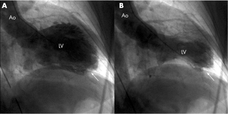
End diastolic (panel A) and end systolic (panel B) ventriculograms of the patient, showing aquinesia of apical segments of the left ventricle (LV) and hypercontraction of basal and mid segments. A striking filling defect highly suggestive of a thrombus was also viewed at the apex (white arrow). Ao, aorta.

An official website of the United States government
Here's how you know
Official websites use .gov
A
.gov website belongs to an official
government organization in the United States.
Secure .gov websites use HTTPS
A lock (
) or https:// means you've safely
connected to the .gov website. Share sensitive
information only on official, secure websites.
