Figure 5.
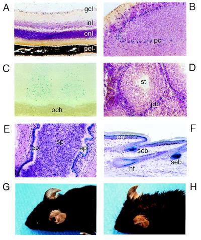
(A–F) Expression of RORα revealed by β-gal activity in RORα+/− mice. Tissue sections were stained with the β-gal substrate 5-bromo-4-chloro-3-indolyl β-d-galactoside (X-Gal) (C) or additionally counterstained with either cresylviolet (A, B, D, and F) or hematoxylin/eosin (E). (A) Retina; gcl, ganglion cells; inl, inner nuclear layer; onl, outer nuclear layer; pel, pigment epithelium layer. (B) Cerebellum; pc, Purkinje cells. (C) Suprachiasmatic nuclei; och, optic chiasm. (D) Testis; ptc, peritubular cells; st, seminiferous tubule. (E) Epididymis; sp, spermatocytes; epi, epithelium. (F) Tail skin; epi, epidermis; seb, sebaceous gland; hf, hair follicle. (G and H) Comparison of a RORα+/− and a RORα−/− mouse; note the difference in the appearance of the fur and the partially exposed skin of the RORα−/− mouse (H).

