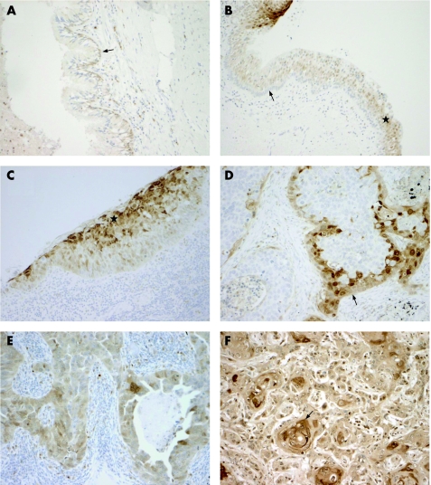
Figure 2 (A) Normal bronchial epithelium is mostly negative for acid cysteine protease inhibitor (ACPI), with only weak expression is seen in the basal cells (arrow). (B) Normal bronchial epithelium adjacent to the dysplastic epithelium. Negative ACPI staining in the basal‐cell layer (arrow) and positive staining in the upper parts of the epithelium (star). The strongly stained dysplastic epithelium is seen in upper left. (C) Dysplastic bronchial epithelium showing strong staining of ACPI in upper two‐thirds of the epithelium (star). (D) Squamous‐cell carcinoma showing a positive staining for ACPI in the peripheral area of the tumour epithelium (arrow). (E) Strong staining for ACPI in adenocarcinoma. (F) Well‐differentiated squamous‐cell carcinoma with strong ACPI staining. Note the concentration of staining signal around the keratin pearls (arrow). Original magnification×330.

An official website of the United States government
Here's how you know
Official websites use .gov
A
.gov website belongs to an official
government organization in the United States.
Secure .gov websites use HTTPS
A lock (
) or https:// means you've safely
connected to the .gov website. Share sensitive
information only on official, secure websites.
