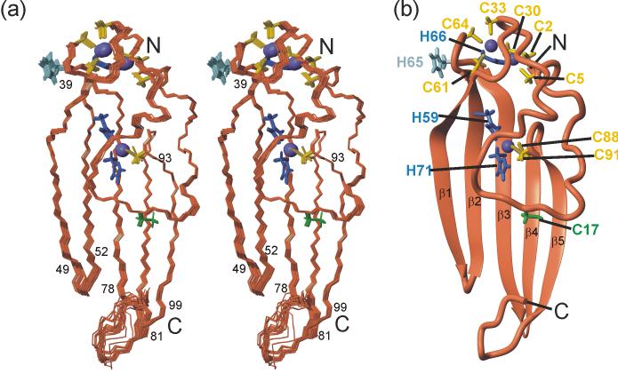
Figure 3.
Solution structure of ChCh107. (a) Stereo representation of a superposition of the heavy atoms for residues 2-99 of the 20 lowest energy structures. Residues at the ends of the β-strands are indicated. (b) Ribbon diagram of the lowest energy refined structure shown in the same orientation with zinc ligands and β-strands labeled. The backbone is shown in coral, zinc in purple, cysteine side chain zinc ligands in yellow, histidine side chain zinc ligands in blue. The unliganded His65 and Cys17 are shown in light blue and green respectively. The figure was prepared with MOLMOL.74

