
Figure 5. Binding of HuC25 and 3D12 FAB fragments to yeast displayed BoNT/A HC alanine mutants.
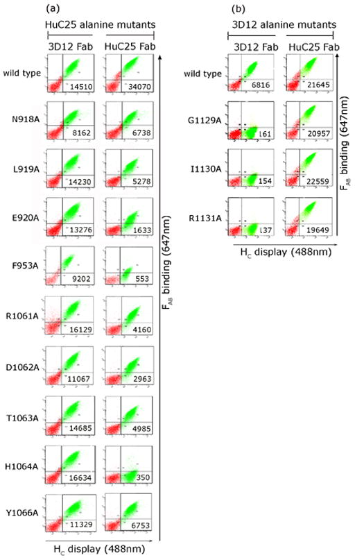
Antibody binding to yeast displayed HC was quantitated on a flow cytometer by co-staining with anti-SV5 (Alexa-488) and FABs HuC25 and 3D12 followed by FAB-specific-APC (647 nm) labeled antisera. Mean fluorescence intensity (MFI) values for FAB binding are shown. Comparison of the MFI for wildtype binding and mutant binding of HuC25 vs 3D12 FAB identified alanine mutations with reduced affinity.