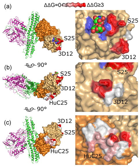
Figure 6. Model of the functional binding epitopes of neutralizing BoNT/A mAbs.

The epitopes of S25 (top panels), 3D12 (middle panels) and HuC25 (bottom panels) are shown, with the ΔΔG of important toxin side chain interactions colored red-white as indicated. The putative sialoganglioside contacting residues on HCC are colored blue, and the sialoganglioside is shown modeled as spheres, near the S25 epitope. HCC, space filling light orange; HCN, space filling dark orange, HN, green, LC, magenta. Molecular models were constructed using Pymol software (DeLano Scientific, LLC), using the coordinates of BoNT/A (3BTA) from the Protein Data Bank.
