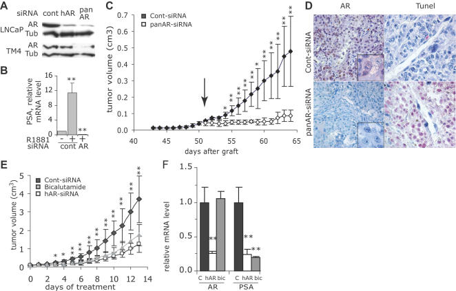
Figure 1. Silencing of AR in LNCaP cells and tumors.
A: Control (cont)- panAR- or hAR-siRNA were transfected into human LNCaP or into mouse Sertoli TM4 cells. AR was immunodetected by western blot in cell lysates 2 days after removal of transfection medium. α-tubulin (tub) expression was used as a loading control. B: Relative PSA mRNA level in LNCaP cells transfected with control or hAR-siRNA and grown for 48 h in the absence of androgens or in the presence of R1881, 0.5 nM (mean±SE, n = 3 independent experiments). Similar results were obtained using the panAR-siRNA. **p<0.01 as compared to values in the absence of androgens. C: LNCaP cells were subcutaneously injected on day 0 to nude mice. Starting from day 51 (arrow), animals (5 per group) received a daily i.p. injection of 3 µg of cont- (black symbols) or panAR-siRNA (white symbols) diluted in 50 µl saline; tumor volume (cm3, mean±SE, n = 5). *p<0.05 and **p<0.01 comparing panAR-siRNA to cont-siRNA treated tumors. D: Analysis of AR expression by immunohistochemistry (left panels) and apoptotic cells by TUNEL (right panels) in representative tumors collected at the end of the experiment shown in C. E: Mice bearing exponentially growing LNCaP tumors were randomized (12 mice per group) and received daily i.p. injections of cont-siRNA (black symbols), hAR-siRNA (white symbols) or an oral dose of 50 mg.kg−1 of bicalutamide (grey symbols); tumor volume (cm3, mean±SE). *p<0.05 and **p<0.01 comparing hAR-siRNA to cont-siRNA treated tumors. F: On the fourth day of treatment of the experiment shown in E, 6 mice in each group were sacrificed and AR and PSA mRNA levels were quantified in the tumors by qRT-PCR and normalized with cyclophilin A mRNA level. Results are expressed relative to the mean level in control tumors. **p<0.01 comparing hAR-siRNA to cont-siRNA treated tumors.

