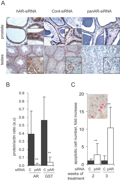
Figure 2. Silencing of AR in prostate and testes.
A: Upper panels, immunodetection of AR expression in the ventral prostate of mice treated for 3 weeks with hAR-, cont-, or panAR-siRNA as indicated. Lower panels, AR expression in testes from mice sacrificed at the end of the experiments shown in figure 1C, after 2 weeks of treatment (cont- and panAR-siRNA) or treated for 3 weeks with hAR-siRNA. B: AR and GST expression in testes from mice treated for 3 weeks with cont- (black bars) or panAR-siRNA (pAR, white bars). AR and GST levels were quantified by immunoblot, normalized with actin level, (arbitrary units, mean±SE, n = 10). C: Quantification of apoptotic germ cells (insert) in testes collected from mice treated for 2 or 3 weeks (mean number/ 100 seminiferous tubules±SE) with cont- (c) or panAR-siRNA (pAR). **p<0.01 as compared to cont-siRNA treated group.

