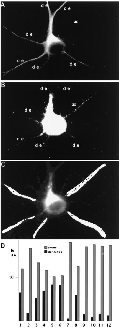
Figure 1.
Axonal distribution of HA in stage 5 neurons. (A) FPV-infected stage 5 neuron stained with an antibody against the dendritic marker MAP2; the axon (ax) appears devoid of labeling. (B) The same infected neuron was coimmunostained with an antibody against HA. The labeling is mainly present in the axon (ax), leaving the dendrites (de) almost unstained. This was quantified by binarizing the image in B and choosing at random similar areas in dendrites and axons as is shown in C. The number of pixels was analyzed and normalized to the same area size in each process. (D) Percentage of HA present in dendrites versus axons in 12 different neurons. The mean value for the distribution of HA is 78% in the axons and 22% in the dendrites.

