
Fig. 3.
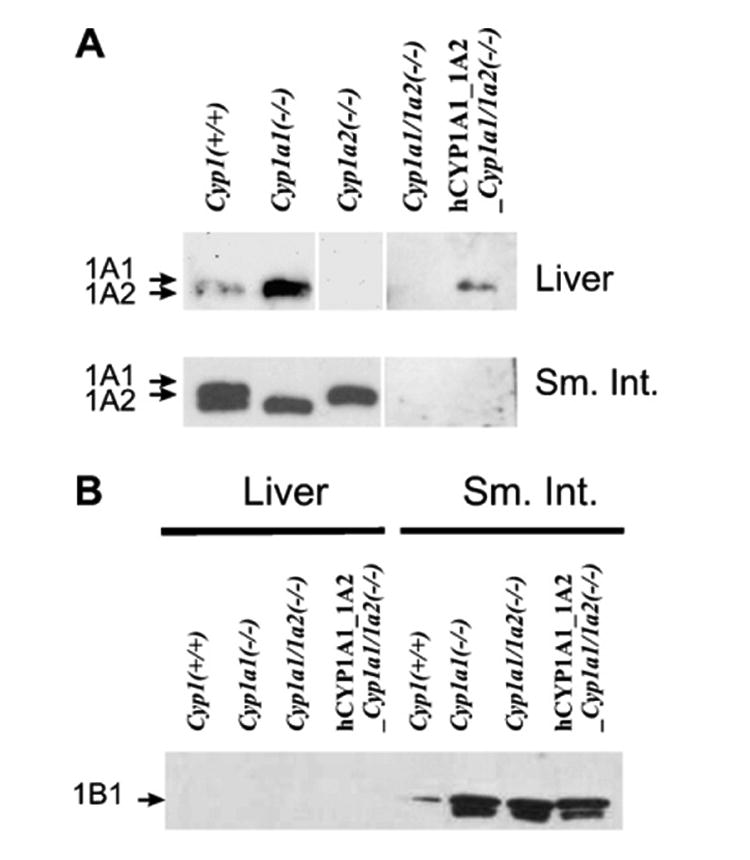
Western immunoblot analysis. (A) Mouse or human CYP1A1 and CYP1A2 proteins in the liver (top row) and proximal small intestine (lower row) from the five genotypes under study. The human CYP1A1 and CYP1A2 proteins migrate between that of the mouse CYP1A1 and CYP1A2 proteins [5]. The same regimen (oral BaP-treated) was employed. The rabbit polyclonal anti-rat CYP1A1/1A2 antibody (Daiichi Pharmaceuticals Co. Ltd., Tokyo) was used; this antibody recognizes both mouse and human CYP1A proteins. (B) Mouse CYP1B1 protein in liver (left) and intestine (right) of the oral BaP-treated mice. Liver and intestine CYP1B1 levels in Cyp1a2(−/−) mice (not shown) exhibited levels similar to that in Cyp1(+/+) mice. A rabbit polyclonal anti-mouse CYP1B1 antibody was developed in our laboratory, in collaboration with Alpha Diagnostic International (San Antonio, TX); this antibody recognizes both mouse and human CYP1B1 protein. Each lane was loaded with 0.25 and 10 μg microsomal proteins for liver and small intestine, respectively. The reason for the faster-moving band in this doublet is not known, but might represent mitochondrial CYP1B1 protein contamination [34] of the small intestine microsomes.